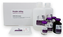
Biocolor’s Purple-Jelley Assay Kit is the perfect tool for accurate measurement of hyaluronic acid / hyaluronan levels in your samples. This colorimetric assay is optimized for quantitative analysis of in-vivo, tissue-derived hyaluronic acid and includes full step-by-step instructions.
Description
The Biocolor Purple-Jelley Hyaluronic Acid Assay is a quantitative, dye-based method for measurement of hyaluronic acid/hyaluronate/hyaluronan derived from tissues.
The assay manual includes methods for the user to remove tissue protein and sulfated glycosaminoglycans before measuring the isolated hyaluronic acid (HA).
Principle of Method
During the assay, hyaluronic acid is selectively purified during the assay sample preparation protocol. This is then reacted with the Purple-Jelley dye reagent (containing ‘Stains-all’ dye), and the absorption of the characteristic third wavelength recorded. By comparison with a calibration curve the hyaluronic acid content of the sample can be measured.
Step 1. The assay protocol takes tissue samples through a sequential sample preparation protocol which involves enzymatic protein digestion, followed by precipitation and purification of GAGs, culminating in the precipitation of purified Hyaluronic acid.
Step2. The processed sample is then incubated for 10 minutes with the Purple-Jelley dye reagent, forming a colored product which can be measured spectrophotometrically.
Step 3. The Hyaluronic acid content of unknown samples can be calculated by comparison against a calibration curve prepared using a standard comprising hyaluronic acid (supplied with the kit).

Sample Type
Tissue extracts.
Removal of tissue protein before assay is essential in order to isolate the HA from potentially dye binding proteins; and sulfated glycosaminoglycans need to be removed before measuring the isolated HA, from a two step critical electrolyte salting out process (CEC). Methods for protein removal and sGAG removal can be found in the product manual.
Specifications
| Product Category: | |
| Detection Method: | Colorimetric Detection (655 nm) |
| Assay Range: | 0 - 100 µg/ml |
| Limit of Detection: | 10 µg/ml |
| Sensitivity: | 0.2 µg |
| Measurements Per Kit: | 100 in total (allows a maximum of 46 samples to be run in duplicate alongside a standard curve) |
| Storage (unopened kit): | Room Temperature (RT) |
| Manufacturer: | Biocolor Ltd. |
| Country of Origin: | United Kingdom |
Kit Contents
The standard size Purple-Jelley™ Hyaluronic Acid kit (cat. no. H1000) contains:
- Purple-Jelley Dye Reagent (1x 20 ml)
- Hyaluronan Reference Standard (1x 5 ml, 0.2 mg/ml soluble Hyaluronic Acid)
- Precipitating Reagent (2x 34 ml)
- Sodium Chloride (1x 20 ml)
- Cetylpyridinium Chloride (1x 20 ml)
- TRIS-buffered Saline (5x tablets)
- 2 ml screw-cap tubes for preparation of samples
- Assay kit manual
Examples of Results
The distribution of HA in tissues as produced in this assay procedure is listed in Table 1 and is given only as an outline guide. In adult animals the HA percentage gradually increases during ageing as muscle and fat mass decreases.

Other Names/Synonyms
Hyaluronic acid, Hyaluronan, HA, Purple-Jelly Hyaluronan Assay
Resources
- Product Datasheet (PDS)
- Material Safety Data Sheet (MSDS)
- Frequently Asked Questions (FAQ)
- Product References (Extended)
2025 Product References
- Singh, Pragya et al. “Comparative Study of Placental Allografts with Distinct Layer Composition.” International journal of molecular sciences vol. 26,7 3406. 5 Apr. 2025, doi:10.3390/ijms26073406
- Ikawa, Tetsuya et al. “Impact of Hyaluronic Acid on the Cutaneous T-Cell Lymphoma Microenvironment: A Novel Anti-Tumor Mechanism of Bexarotene.” Cancers vol. 17,2 324. 20 Jan. 2025, doi:10.3390/cancers17020324
Usage
This product is intended for laboratory research use only and is not for use in diagnostic procedures.
Related Products
- Extracellular Matrix Assays
- Proteinase K
- 3-D Life PVA-HA Hydrogel Kit
- 3-D Life Dextran-HA Hydrogel Kit
-
Hyaluronidase Enzyme (Bovine purified)
Background
Hyaluronic acid, in its hydrated form, is a unique polysaccharide polymer, often referred to as a 'gentle giant'. It is found in many tissues, but particularly in cartilage and synovial fluid and consists of a lengthy, flexible, non-branching chain with a repeating disaccharide pattern. This pattern comprises alternating uronic acid and aminosugar units. Hyaluronic acid is involved in a wide range of roles including tissue hydration and lubrication, cell signaling, extracellular matrix reorganization and wound repair.
This involvement of Hyaluronic acid in tissue lubrication and wound healing has meant it has become a well-known additive to both medical and cosmetic skincare treatments, as well as treatments for osteoarthritis. The ability to assay or measure hyaluronic acid is therefore of interest to scientific researchers from a range of disciplines.
Ilex Life Sciences LLC is an official distributor of Biocolor products.


