Bring natural bone environments to the laboratory
What are P3D Scaffolds?
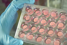
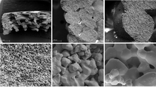
Ossiform P3D Scaffolds are xeno-free, bioceramic scaffolds which provide a bone-like niche for in vitro and in vivo studies such as disease modeling, drug screening and bioengineering. The 3D printed bioceramic scaffolds are developed to resemble native bone with respect to both physical and structural properties, such as high contents of β-tricalcium phosphate (β-TCP) and stiffness, as well as macro- and micro-porosities.
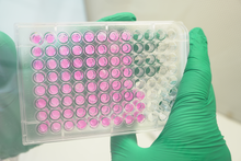
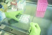
The P3D Scaffolds are an easy plug-and-play solution for 3D cell culturing of bone harboring or bone related cell types. The cell culture system is compatible with most common laboratory techniques and does not require major alterations of preexisting protocols and workflows.
P3D Scaffolds provide you with a clinically relevant cell culture system of natural materials and customized structures which allow you to create predictive research models of human physiology and pathology.
Advantages
- Enable complex co-culturing
- Maintain relevant cell signaling
- Support the natural self-organization of cells
- Easily apply 2D analysis methods
- For in vitro and in vivo pre-clinical research
- All P3D Scaffolds are supplied sterile and ready to use
Benefits
- 100% polymer-free
- Consist solely of β-tricalcium phosphate (β-TCP)
- Clinically relevant environment
- Directly translate from in vitro to in vivo
- Fully customizable to your research needs
- Easy-to-use 3D cell culture and drug screening systems
- Animal-free alternative to bone slices
Applications
The P3D Scaffold enables the creation of good disease models, such as bone tumors, and is useful for studying diseases like bone metastasis, osteomyelitis, and osteoporosis. Various cells and pharmaceuticals can be added to the P3D Scaffolds to study how they interact with each other and with the bone within its pores. Potential usages include, but are not limited to, the examples mentioned in the following:
- Mesenchymal stem cells (MSCs) or osteoblasts can be added onto the P3D Scaffold to study how new bone develops in bone grafts.
- Bone destruction in osteoporosis/arthritis can be imitated by seeding osteoclasts and macrophages onto the scaffolds.
- Resorption assays with human osteoclasts for studying osteoclast activation and resorption in various culturing conditions.
- The development of bone tumors and spreading of cancerous cells through the outer calcified bone matrix can be studied by seeding cancerous cells onto the P3D Scaffolds.
- Osteomyelitis and the subsequent destruction of bone can be mimicked by adding bacteria and immune cells to the P3D Scaffolds. This enables you to study the mechanisms behind the bacteria's destruction of the hardened calcified bone as well as how bacteria are able to evade the immune system and antimicrobial pharmaceuticals.
Many methods may be used for analyzing the biological activity on the 3D printed structures. Visit our P3D Scaffolds FAQ for more information.

Explore P3D Scaffolds - Standard Design Options
Choose Design
Gyroid or grid

Different infill types fulfill your different research needs.
Do you need to monitor cell morphology close while the cells are still in culture? Then go for the grid structure, which allows light to pass through the scaffold easily, thereby enabling you to perform regular checkups on your cells. Our data shows that cells will grow from the corners and in.
Is your research more focused on gaining a model system as natural as possible? Then the gyroid structure is just the right choice for you. Here you will get a randomized organization of the pores, resulting in a variable pore size, making regular checkups while in culture a bit trickier, but leaving you with a more accurate representation of the bone structure.
Choose Porosity
Choose between:
- Porosity 1: 300-400 micrometers
- Porosity 2: 600-800 micrometers (not recommended for 5 mm diameter scaffolds, as it effects the macro porosity due to the small scaffold size).
Choose Size
Four standard scaffold diameters are available, designed to fit common cell culture plate sizes. All standard P3D Scaffolds have a height of 3 mm.
- 5 mm diameter: suitable for 96-well plates
- 12 mm diameter: suitable for 24-well plates
- 20 mm diameter: suitable for 12-well plates
- 30 mm diameter: suitable for 6-well plates

Extra customizable features
Special forms, sizes (height, diameter, etc.), and designs of the scaffolds can be requested for a fully customized set of cell culture systems. Please contact us to discuss your specific needs.
Resources
- P3D Scaffolds Product Data Sheet (PDS)
- P3D Scaffolds Product Brochure
- P3D Scaffolds Frequently Asked Questions (FAQ)
- Scaffolds-Based 3D Cell Culturing Research Resource Handbook
- P3D Scaffolds Material Safety Data Sheet (MSDS)
- Protocol: P3D Scaffold Cell Recovery by Trypsinization
- This protocol explains how to retrieve cells that have been cultured on the P3D Scaffolds. Cell recovery from the P3D Scaffolds can be performed using standard protocols with minor modifications.
- Protocol: How to Culture hMSCs in Bioceramic 3D Cell Culture Systems
- This protocol is for seeding and 3D culturing human mesenchymal stem cells (hMSCs) on P3D Scaffolds. The procedure provides guidelines on how to create three-dimensional multicellular in vitro tissue constructs based on hMSCs and subsequently differentiate the hMSCs into osteoblasts to create a 3D bone cell culture.
- Protocol: How to Verify Osteogenesis on P3D Scaffolds
- This guide is for studying osteoblast differentiation using an ALP staining assay. Mesenchymal stem cells express alkaline phosphatase (ALP), which increases during osteoblast differentiation. ALP levels can therefore be used as an osteogenic marker.
-
Protocol: Evaluation of Cell Viability Using CellTiter-Glo 3D Cell Viability Assay with P3D Scaffolds
- This protocol explains how to evaluate cell viability using CellTiter-Glo 3D cell viability assay with Ossiform P3D Scaffolds. This assay lyses all cells and measures the amount of ATP present. It works by mixing cells in media 1:1 with Assay Reagent and measuring output luminescence.
-
Protocol: Evaluating Cell Viability Using Invitrogen LIVE/DEAD Cell Imaging Kit with P3D Scaffolds
- This protocol explains how to perform cell viability using Invitrogen LIVE/DEAD Cell Imaging Kit with Ossiform P3D Scaffolds.
-
Protocol: Protein Extraction with Ossiform P3D Scaffolds
- A protocol for harvesting protein from cells cultured on P3D Scaffolds.
- Protocol: Harvesting RNA from Cells Cultured on P3D Scaffolds
- This protocol explains how to easily recover RNA from cells cultured on P3D Scaffolds.
Product References
- Gomes, G. H. M., J. K. M. B. Daguano, J. G. Bastidas, K. N. Ferreira, P. H. J. O. Limirio, P. . Dechichi, and L. E. . Carneiro-Campos. “Characterization of 3D-Printed B-TCP Scaffolds With Enhanced Microstructure, Mechanical Properties, and Cell Compatibility”. International Journal of Advances in Medical Biotechnology - IJAMB, vol. 6, no. 2, Dec. 2024, pp. 61-76, doi:10.52466/ijamb.v6i2.131.
Ilex Life Sciences is an official distributor of Ossiform P3D Scaffolds research product line.


