
Protocol: Evaluation of Cell Viability Using CellTiter-Glo 3D Cell Viability Assay with P3D Scaffolds
Application
This protocol explains how to evaluate cell viability using CellTiter-Glo 3D cell viability assay with Ossiform P3D Scaffolds.
This assay lyses all cells and measures the amount of ATP present. It works by mixing cells in media 1:1 with Assay Reagent and measuring output luminescence. The output requires the use of black/opaque 96-well plates with clear bottoms. This procedure describes how to use this kit with scaffolds. Please also refer to manufacturer Promega’s protocol.
| Plate type | Media volume | Assay Reagent volume |
| 96-well plate | 100 uL | 100 uL |
| 24-well plate | 200 uL | 200 uL |
| 12-well plate | 500 uL | 500 uL |
| 6-well plate | 1500 uL | 1500 uL |
Materials
- CellTiter-Glo 3D cell viability assay
- Plate shaker
- Opaque plate
- Plate reader
Flowchart

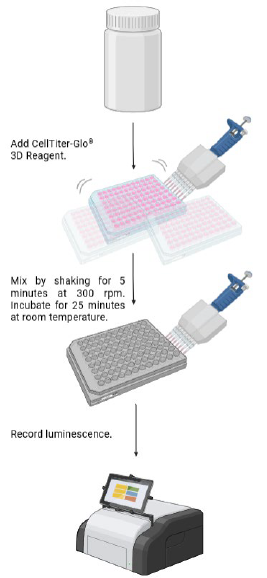
Figure 1.: Workflow for evaluating cell viability using CellTiter-Glo 3D cell viability assay.
Notes before starting and general advice on material handling
- All handling of the P3D Scaffolds products should be performed using gloves, according to the standard aseptic methods.
- The scaffolds are supplied sterile by dry heat sterilization and remains sterile until opened.
Procedure
The day before performing the procedure: place Assay Reagent at 4C to allow for slow thawing.
Before you start:
- Place Assay Reagent and cell plates at RT to equilibrate for at least 30 min.
- Gently invert assay reagent bottle to mix before use.
- Make aliquot of assay reagent by calculating the amount needed.
- If necessary, remove media from wells to achieve volumes listed in table above.
Procedure:
- Add Assay Reagent to each well, using multichannel pipette if possible. Volume of Assay Reagent should be equal to the volume media already present in wells.
- Place plate at RT on a plate shaker at 300 rpms for 5 min.
- Remove plate from plate shaker and incubate plate at RT for 25 min.
- Pipette 200 uL lysate to opaque plate as quickly as possible.
- Measure luminescence using a plate reader with integration time 0,25-1 seconds. Settings may vary between instruments.
Disclaimer
The products are “For Research Use Only (RUO)” and should not be used for clinical, diagnostic, or therapeutic purposes. Ossiform and its distributor, Ilex Life Sciences LLC, make no other warranties, expressed or implied, including the implied warranty of merchantability and the implied warranty of fitness for particular purpose.
Acknowledgement
This protocol has been adapted from the protocol published by Ossiform. Ilex Life Sciences is an authorized distributor of Ossiform P3D Scaffolds research product line.