Introduction
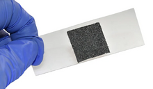
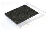
3D G-Scaffold is a xeno-free, highly porous, conductive carbon scaffold designed for 3D cell culture, tissue engineering, and regenerative medicine applications. Engineered using proprietary chemical vapor deposition (CVD) technology, 3D G-Scaffolds offer a consistent 3D nanofibrous network mimicking the native extracellular matrix (ECM) with a large surface area for cell attachment and growth.
With an interconnected microporous network composed of conductive graphene sheets, the 3D G-Scaffold provides ideal biomechanical, biochemical, and electrical properties to grow healthy stem cells than maintain stemness and provide more relevant experimental outcomes.
The 3D G-Scaffold has been tested with a variety of cells - including fibroblasts, induced pluripotent stem cells (iPSCs), and mesenchymal stem cells (MSCs) - across several tissues - including cardiovascular and bone cells.
Cells grown on 3D G-Scaffold exhibit a high degree of attachment, maintain their phenotype, and grow as an interconnected network. The 3D G-Scaffold has also demonstrated improved gene expression compared to 2D cell culture plastic and glass substrates.
When compared to other commercially available scaffolds, 3D G-Scaffold has shown improved cytocompatibility, cell adhesion, cell morphology, and cell viability.
Features
- Complex co-culturing of cells.
- Supports application of electric field to guide cell proliferation, differentiation, and maturation.
- Surface treatment or protein coatings not required to improve cell adhesion.
- For in vitro and in vivo pre-clinical studies.
- Clinically relevant environment.
- Composed solely of carbon.
- Proven biocompatibility.
- Supports cell imaging using standard staining protocols for fluorescence or confocal microscopy.
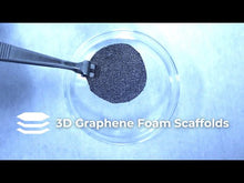
- Safe to cut with scissors and handle lightly with tweezers at the edge.
Applications
Graphene foam biomaterial based 3D G-Scaffolds are suitable for a broad range of research applications. For example:
-
Conductivity and Electrical Field Application
- 3D G-Scaffolds are highly conductive – making them a novel and unique platform to study how electrical stimulation via external and direct field application can influence stem cell behavior. It has been found that 3D G-Scaffolds in combination with electrical stimulation can guide stem cell differentiation, accelerate maturation and growth, and support overall tissue regeneration across bone, cardiac, neural, cartilage, and several other tissue types.
-
Wound Healing
- 3D G-Scaffolds in combination with mesenchymal stem cells (MSCs) have been found to deliver the necessary bioactive cues for enhanced and rapid wound healing - with enhanced vascularization and reduced scarring. It has also been found that the excellent biocompatibility of 3D G-Scaffolds loaded with MSCs permits excellent integration with the host tissue.
-
Neural Tissue Engineering
- 3D G-Scaffolds have been seeded with a variety of neural stem cells (NSCs) and have demonstrated the ability to maintain NSCs in an active proliferation state with noticeable upregulation of protein expression. Phenotypic analysis confirms that 3D G-Scaffolds can enhance NSC differentiation towards astrocytes and especially neurons, and can reduce inflammation from external factors. 3D G-Scaffolds can also be utilized to develop customized conductive scaffolds for neural-machine interfaces or neuro-prosthetic devices.
-
Cardiovascular Tissue Engineering
- 3D G-Scaffolds offer an electrically active interface to culture cardiomyocytes while being able to directly monitor their extracellular activity. With a highly porous interconnected network, nano-micro scale topographical surface, and a highly conductive interface, 3D G-Scaffolds demonstrate robust attachment of both cardiomyocytes and cardiac fibroblasts without any cell adhesion coating - with no adverse effects on cell viability.
Specifications
| Growth Method: | Chemical Vapor Deposition (CVD) |
| Quality Control: | Raman, SEM, and Optical |
| Growth Substrate: | Nickel Foam |
|
Format: |
Graphene foam insert |
| Appearance: | Freestanding foam |
| Dimensions (cm): | 3 x 3 x 0.16 cm |
| Composition (EDS): | > 99% C |
| Density (mg/cm3): | 27 +/- 6 mg/cm3 |
| Surface Area (m2/g): |
~ 400 m2/g [1] [1] DOI 10.1088/2053-1591/ab2e83 |
| Pore Size (μm, average): | 5 - 500 μm |
| Conductivity (S/m): | 2 x 10^5 S/m |
| Yield Stress @ 0.5% Strain (kPa) | 3.7 +/- 2 |
| Compressive Modulus (kPa) | 120 +/- 80 |
| Supported Sterilization Protocols: | 70% ethanol, UV, autoclave compatible |
| Storage: | Room temperature |
| Manufacturer: | General Graphene Corporation |
Conductivity
With its highly conductive structure, the 3D G-Scaffold supports the use of external electric fields for in-depth studies on specific cell behaviors. This method offers a dynamic way to investigate the effect of electric stimulation on cells, enhancing our understanding of neural activity, cardiac function, and drug action mechanisms.

Cardiac fibroblasts adhered to 3D graphene foam.
Biomechanical Properties
The biomechanical properties of 3D G-Scaffold - specifically its interconnected microporosity and nanoroughness - play a role in mediating a cell's ability to sense its extracellular environment, enhancing spatiotemporal and biological cell behavior.

Lonza Mesenchymal Stem Cells (MSCs) are evenly distributed across the graphene foam and are mostly found wrapping around the pores. Captured data on volume and sphericity shows cells spreading.

Lonza Mesenchymal Stem Cells (MSCs) demonstrate an elongated morphology while forming a mesh of interconnected cells adhered to the graphene foam.
Cell Attachment
The 3D G-Scaffold eliminates the need for surface treatment or coating with biological materials, such as gelatin, poly L-Lysine, or Matrigel which can interfere with experimental results. This feature ensures that results are free from external influences during in vitro and in vivo experiments - leading to more reproducible outcomes without concerns over damage caused by the surface treatment or coating application.

Fibroblast cells were cultured for 24 hours on the graphene foam, demonstrating an elongated morphology without requiring an ECM protein coating. Actin cytoskeleton is stained with phalloidin (orange) and the nucleus with DAPI (light blue).
Cell Viability
3D G-Scaffold is biocompatible and demonstrates negligible toxicity to MSCs.

Live-Dead Assay performed using Lonza Mesenchymal Stem Cells (MSCs) showed significantly higher number of live cells - with only a 0.6% decrease in the number of live cells from Day 3 to Day 7.
Documents
- Product Data Sheet (PDS)
- Material Safety Data Sheet (MSDS)
- Guidelines for Cell Culture Using 3D G-Scaffold
- 3D G-Scaffold Handling Guide
- Protocol: Cell Seeding Protocol for 3D G-Scaffold
- Protocol: Cell Dissociation Protocol for 3D G-Scaffold
Customization
3D graphene foam can be custom produced in the ideal sizes and formats to meet your specific research and handling requirements. For example, 3D G-Scaffolds can be provided in a 6-well culture plate, as seen below. Matrix materials can be embedded in the scaffolds on request. Please contact us to discuss your specific needs.

Ilex Life Sciences is an authorized distributor of General Graphene Corporation products.


